Vibhuti PathakBy: Vibhuti Pathak
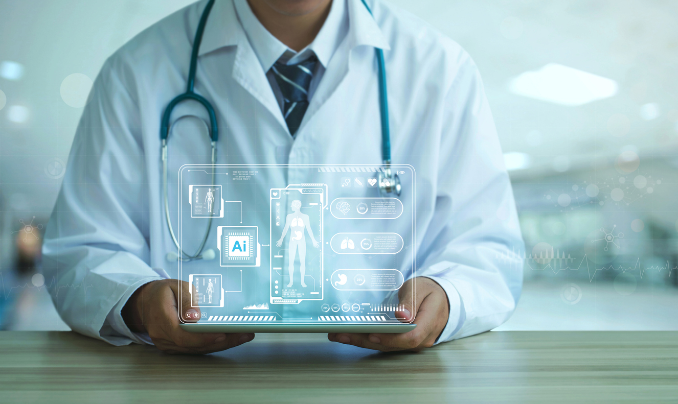
Researchers have introduced an innovative artificial intelligence (AI) model capable of precisely identifying tumours and diseases in medical images.
Published in the journal IEEE Transactions on Medical Imaging, this unique AI model provides transparency by explaining each diagnosis through a visual map. This transparency enables doctors to easily comprehend the reasoning behind the AI’s decision, double-check for accuracy, and communicate results effectively to patients.
“The idea is to help catch cancer and disease in its earliest stages — like an X on a map — and understand how the decision was made. Our model will help streamline that process and make it easier on doctors and patients alike,” said Sengupta, the study’s lead author,” said Sourya Sengupta, a graduate student at Beckman Institute for Advanced Science and Technology in the US.
Medical imaging interpretation varies across the globe, particularly in developing countries facing a shortage of doctors and long patient queues. Sengupta highlights the potential of AI in such scenarios, suggesting that automated medical image screening can serve as an assistive tool, supplementing the expertise of doctors rather than replacing it.
In situations where time and medical expertise are scarce, automated screening can act as a valuable assistive tool. The AI model can pre-scan medical images, flagging those with anomalies, such as tumours or early signs of diseases. This approach not only saves time but can enhance the performance of medical professionals responsible for reviewing the scans.
“In many developing countries, there is a scarcity of doctors and a long line of patients. AI can be helpful in these scenarios,” added Sengupta.
While existing AI models often categorise images as “tumour versus non-tumour,” this new AI model takes a step further. It interprets itself with each decision, explaining its diagnosis rather than offering a binary classification. This self-interpretation feature addresses the common query of patients asking why an AI flagged a particular image.
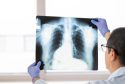
The researchers trained the model on three distinct disease diagnosis tasks, involving over 20,000 images. The tasks included reviewing simulated mammograms, analysing optical coherence tomography (OCT) images of the retina, and studying chest X-rays.
The model’s accuracy rates were impressive, reaching 77.8 per cent for mammograms, 99.1 per cent for retinal OCT images, and 83 per cent for chest X-rays. These high accuracy rates are attributed to the deep neural network of the AI, which mimics the nuanced decision-making process of human neurons.
This groundbreaking AI model not only enhances accuracy in disease diagnosis but also prioritises transparency and interpretability, making it a valuable tool in the field of medical imaging.